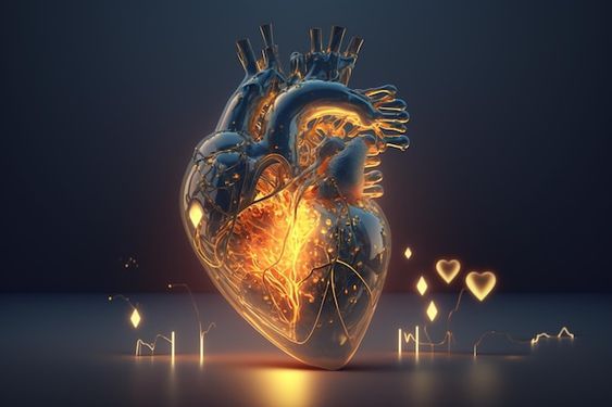
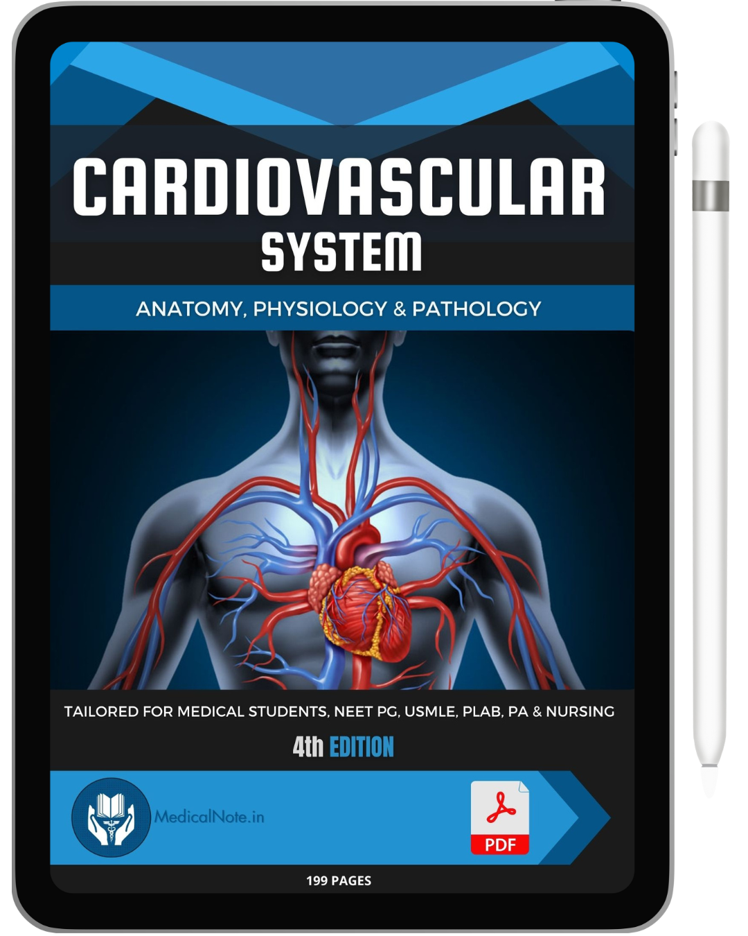
The human heart is an incredible organ, tirelessly working to pump blood throughout the body, delivering essential nutrients and oxygen to tissues. Understanding the heart's anatomy is fundamental to grasping how this vital organ functions and maintains our overall health. In this blog post, we will explore the intricate anatomy of the heart, including its location, structure, and the critical role it plays in the cardiovascular system.
Anatomical Location: The heart is located in the middle mediastinum, nestled between the lungs, slightly left of the midline. This central location is strategic, allowing the heart to efficiently pump blood to the entire body. Surrounded by the pericardium, a double-walled sac, the heart is well-protected and anchored within the chest cavity. The pericardium not only secures the heart but also prevents overexpansion, ensuring it functions within optimal parameters.
Layers of the Heart: The heart wall consists of three distinct layers, each playing a crucial role in its function:
- Epicardium: The outermost layer, the epicardium, serves as a protective layer and is part of the pericardium. It contains fat and connective tissue, providing a smooth surface that reduces friction as the heart beats.
- Myocardium: The middle layer, the myocardium, is composed of cardiac muscle. This muscular layer is responsible for the heart's contractile force, enabling it to pump blood effectively. The myocardium is thickest in the ventricles, particularly the left ventricle, which pumps blood throughout the entire body.
- Endocardium: The innermost layer, the endocardium, lines the heart's chambers and valves. This smooth, thin layer ensures that blood flows smoothly within the heart, reducing the risk of clot formation.
Fibrous Skeleton of the Heart: The fibrous skeleton of the heart is a network of dense connective tissue that supports the heart's structure and function. It anchors the cardiac muscles and valves, maintaining the integrity of the heart's shape and facilitating efficient contraction. Additionally, the fibrous skeleton electrically isolates the atria from the ventricles, ensuring coordinated contractions and proper blood flow.
Heart Chambers and Circulation: The heart comprises four chambers: two atria (upper chambers) and two ventricles (lower chambers). Blood flow through the heart is a well-orchestrated process:
- Right Atrium: Receives deoxygenated blood from the body through the superior and inferior vena cava.
- Right Ventricle: Pumps deoxygenated blood to the lungs via the pulmonary artery for oxygenation.
- Left Atrium: Receives oxygenated blood from the lungs through the pulmonary veins.
- Left Ventricle: Pumps oxygenated blood to the body through the aorta, delivering oxygen and nutrients to tissues.
Heart Valves: The heart has four valves that ensure unidirectional blood flow:
- Tricuspid Valve: Located between the right atrium and right ventricle.
- Pulmonary Valve: Between the right ventricle and the pulmonary artery.
- Mitral Valve: Between the left atrium and left ventricle.
- Aortic Valve: Between the left ventricle and the aorta.
These valves open and close in response to pressure changes within the heart, preventing back flow and ensuring efficient circulation.
Conclusion: The heart's anatomy is a marvel of biological engineering, designed to sustain life by ensuring efficient blood circulation. Understanding its structure and function is essential for appreciating the complexity and importance of this vital organ. Regular cardiovascular health check-ups and a heart-healthy lifestyle can help maintain the heart's function, contributing to overall well-being.


